Can AI Check a Photo of Your Pet's Injury? What a Triage Scanner Sees
A good photo can show things a description can't. It still can't show everything, and knowing where that line sits is what makes photo triage useful.
 Most people describe an injury as "red", "swollen" or "a bit gross". A photo gives the AI the colour, size and surface of the wound to analyse, not just the words.
You notice a red patch on your dog's flank while you're brushing them. Or your cat has a lump near the base of the tail that you're fairly sure wasn't there last week. Or there's a small cut on a paw pad after a walk and you can't tell whether it needs stitches or a wipe with saline.
Your first job is to put what you're seeing into words, and that's where things usually go wrong. "It's red and a bit swollen" could describe a mild insect bite or a fast-spreading hot spot. Size, colour, discharge and edges are hard to describe accurately when you're worried and your pet won't stay still.
That's why PetCare AI has added the Triage Scanner: take a photo of the injury, rash or swelling, and get a clear urgency level with next steps. This post covers how it works, how to take a photo it can use, and where a photo stops being enough.
Why a photo changes the assessment
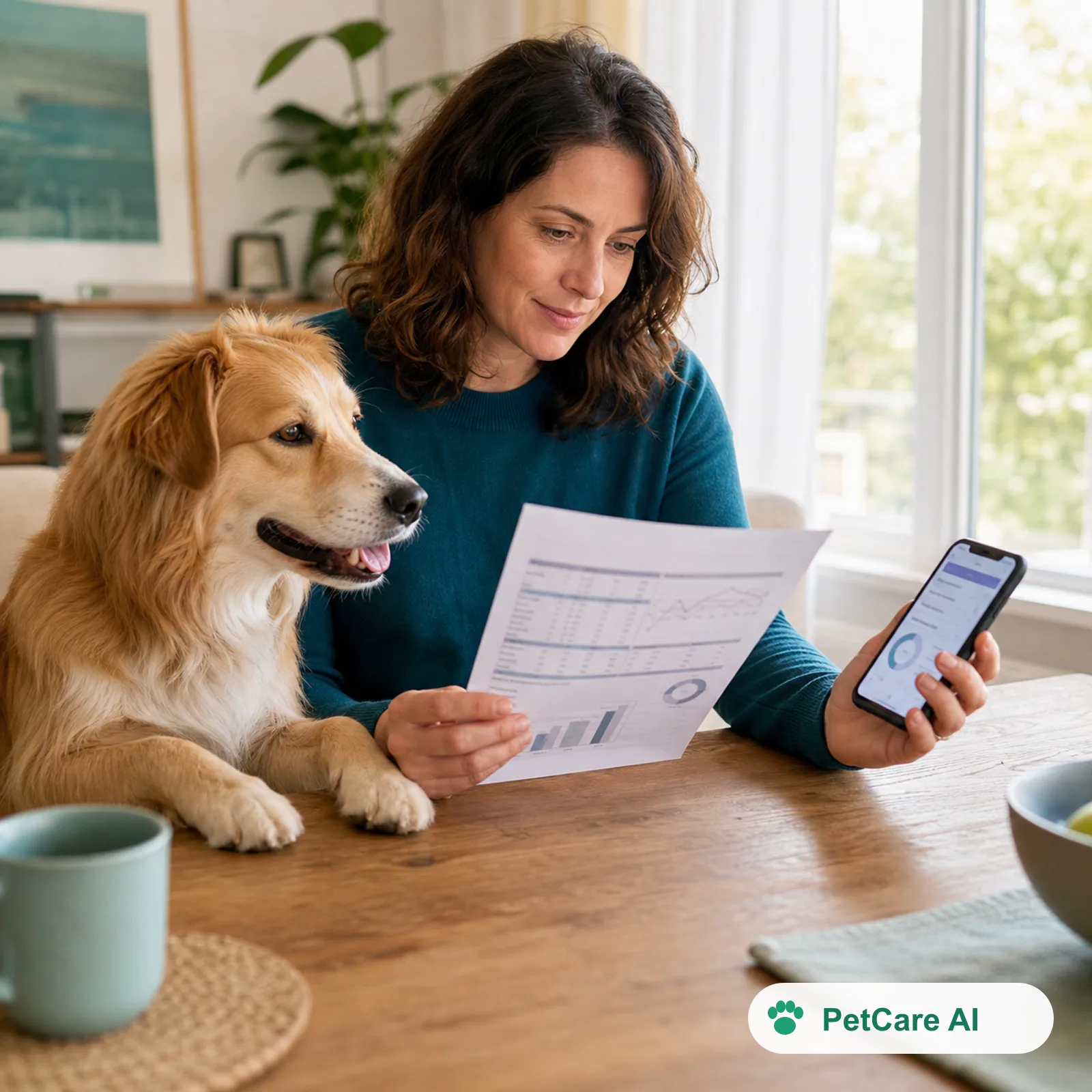
Vets have long used photos to make remote advice better. In a 2026 retrospective study of 100 canine and feline eye emergencies triaged by email, veterinary ophthalmologists correctly identified ocular pain in 79% of cases overall. When a photo was attached, that rose to 89%.1 Agreement between the remote diagnosis and the in-person diagnosis was strong across eye conditions as a whole.1
Those were specialists reading the photos, not an AI. But the result still matters here: the photo added information the written description didn't have.
The same study also found a limit. For corneal conditions specifically, remote agreement with the in-clinic diagnosis was unsatisfactory.1 Some problems can only be judged properly with an examination. We come back to this in the limitations section below.
How the Triage Scanner works
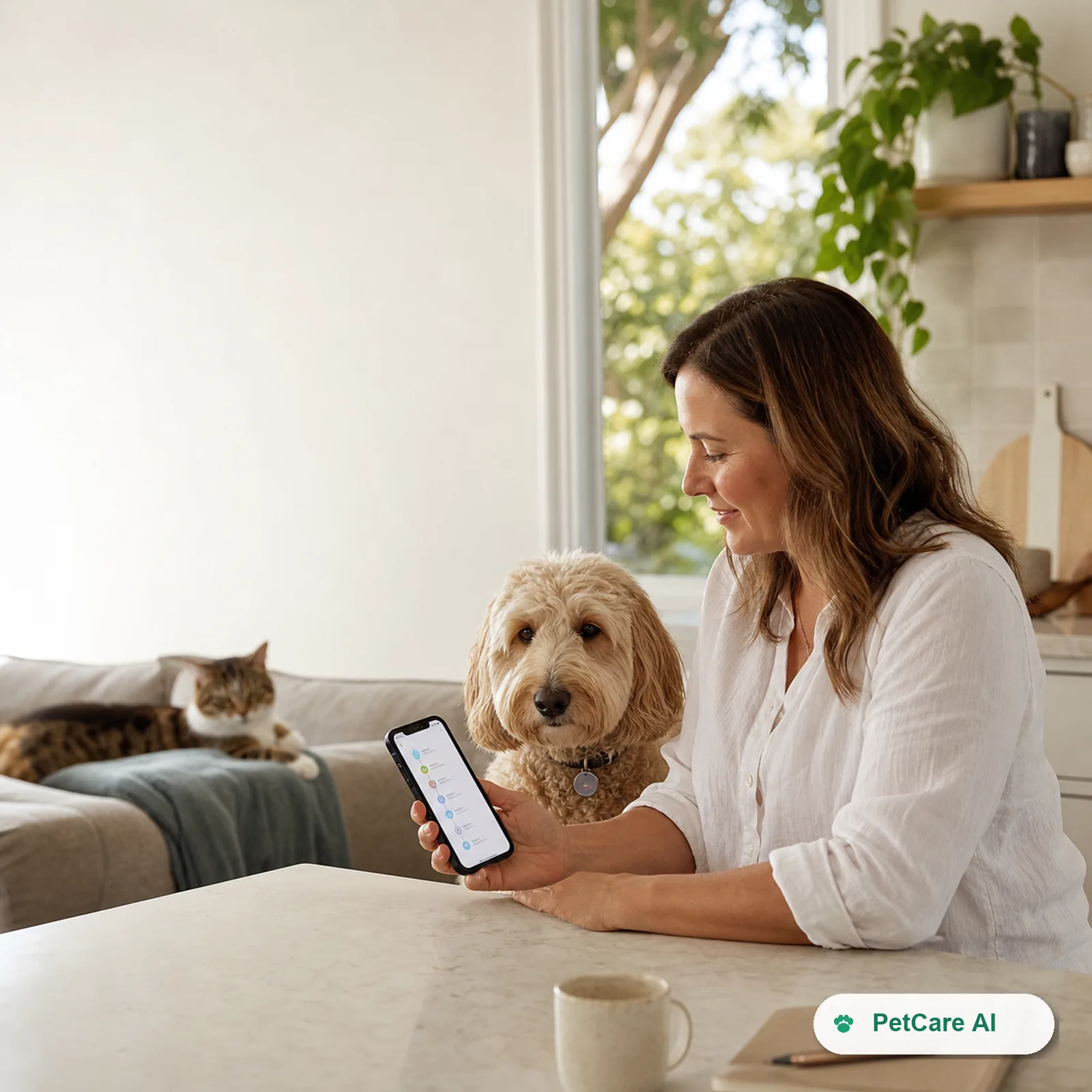
The Triage Scanner is a three-step flow:
- Snap a clear photo of the affected area: a wound, rash, lump, swelling or sore spot.
- The AI analyses the image: it maps the visible issues and assesses them against veterinary triage protocols.
- You get triage steps straight away: an urgency level with the reasoning behind it.
Most of the work happens in step two.
What the AI actually does with your photo
A worried owner looking at a wound tends to see one thing: something's wrong. The AI looks at the image in a structured way. It breaks the photo into the specific visual features that vets use to judge how urgent a skin or surface problem is:
| What the AI looks at | Why it matters for urgency |
|---|---|
| Colour: pink, bright red, dark red, purple, yellow or black | Can separate mild irritation from inflammation, bruising, infection or damaged tissue |
| Swelling: raised, puffy or distorted compared with the surrounding area | Shows how much the area is reacting, and whether swelling is near the airway |
| Surface: intact skin, graze, open wound, puncture, scab | Broken skin raises the risk of infection and changes the first-aid advice |
| Moisture and discharge: dry, weeping, crusted, pus | Wet, oozing patches often point to active infection or a hot spot |
| Size and edges: small and defined, or large and spreading | Spreading or poorly defined edges are more concerning than a stable spot |
| Location: eye, face, paw, joint, belly, near the tail | The same wound means something different on an eyelid than on a flank |
The AI doesn't stop at listing features. It reads them together. A small red bump on a flank is one thing. The same colour with swelling, a wet surface and fast-spreading edges is a different picture. Combining several visual signs into one urgency judgement is the part people find hardest to do in a stressful moment, and it's where AI helps most.
The AI then checks that combined picture against veterinary triage protocols. These are the same structured rules clinics use to decide who is seen first. It returns an urgency level with its reasoning, so you can see why the photo was rated the way it was rather than getting a bare verdict.
Two things are worth keeping in mind when you read an AI image assessment:
- It can only assess what's visible. Image analysis works on the pixels in front of it. It sees the surface of the skin, not what's underneath (more on this below).
- The photo limits the analysis. Blur, glare or fur over the area hides the features the AI relies on. If you're not confident the photo shows the problem clearly, take a better one. That's why photo quality matters so much (see below).
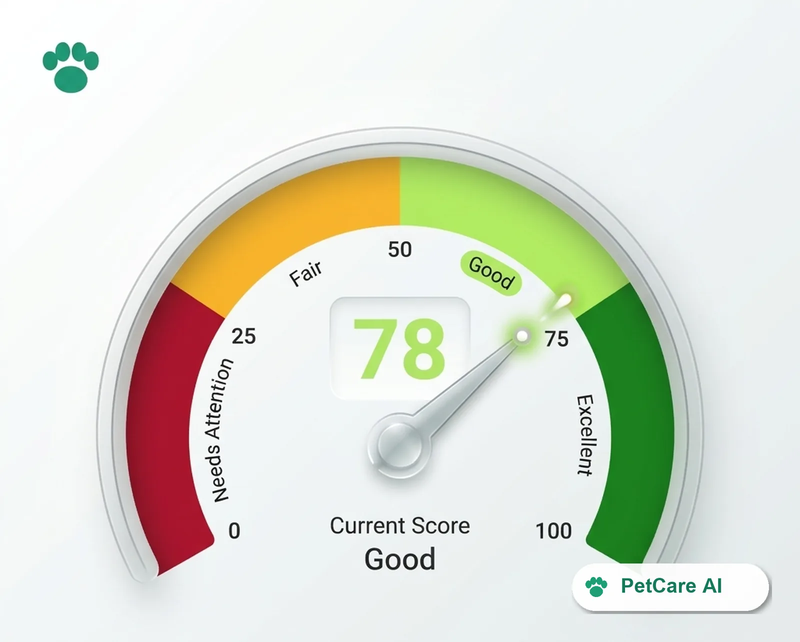
Every result uses the same four urgency levels as the rest of PetCare AI:
| Urgency | What it generally means |
|---|---|
| Emergency | Go to a vet now. Call ahead from the car. |
| High | Contact your vet today, as soon as possible. |
| Moderate | Arrange a vet visit soon, and monitor closely in the meantime. |
| Low | Usually OK to manage at home and monitor, with clear signs that would change that. |
Each level comes with an explanation, what to do right now, and the warning signs to watch for. That last part is often the most useful: it tells you exactly what would turn a "monitor at home" result into a trip to the vet.
A worked example
An illustrative example with a fictional pet.
Bella is a five-year-old Golden Retriever. On Saturday morning her owner notices a small pink patch below her ear that she keeps scratching. By late afternoon it's wet, red, about the size of a palm, and the fur around it is matted.
That pattern fits a hot spot (acute moist dermatitis): a red, inflamed, oozing patch of skin that dogs make worse by licking and scratching. VCA's vets describe owners noticing a small red area before work and coming home to "a raw lesion the size of a pancake".2 Fleas, allergies, ear infections and poor grooming are common triggers.2
Described in words, "a red itchy patch" sounds minor. From the photo, the AI can see what the words leave out: bright red colour, a wet, weeping surface, matted fur at the edges, and a patch much larger than a single bite. Taken together, those features are what should move the result from Low to Moderate. The next steps would include:
- Now: stop the licking and scratching, keep the area clean and dry, and don't apply human creams
- Book: a vet visit, because hot spots usually need clipping, cleaning and treatment of the underlying trigger
- Watch for: the patch spreading, a bad smell, pus, Bella becoming flat or off her food, or a hot spot near the ear alongside head-shaking (a possible ear infection)
The AI didn't diagnose anything. Its image analysis made the urgency reading match what was actually on Bella's skin, not the owner's first description of it.
How to take a photo the AI can read well
AI image analysis is only as good as the image. Colour, swelling, surface and edges all need to be visible for the AI to assess them. The ophthalmology study above found that photo quality was linked to how often the remote assessment matched the in-person one.1 A few habits make a big difference:
- Use natural light, without harsh flash. Flash washes out redness and can hide how deep a wound is. A window or daylight works best.
- Part the fur. On long-coated pets, hold the hair back so the skin is visible, not just the top of the coat.
- Take one close-up and one wider shot. The close-up shows detail. The wider shot shows where on the body it is and how big it is compared with the rest of your pet.
- Add something for scale. A coin or your fingertip beside the area shows whether a lump is 5 mm or 5 cm.
- Keep it steady and in focus. Tap the screen to focus on the injury, not the background.
- Photograph it again later. A second photo a few hours later tells you whether it's spreading, and spreading is often the thing that matters.
 Same graze, two photos. On the left, the AI can barely make out the colour or edges. On the right, it can see the colour, the surface and the size, measured against a 20c coin.
Honest limitations: what a photo can't see
This section matters more than any other in the post.
A photo can't show pain, depth or what's happening inside the body. Merck's veterinary guidance notes that bite wounds can hide serious injuries, such as damaged tissue, broken ribs or injured organs, even when the marks on the surface look minor.3 A cat bite puncture can look like a tiny scab and still become an abscess days later. A photo shows the surface, and the surface isn't the whole injury.
It can't show breathing, behaviour or neurological signs. In Australia this matters. A dog with a paralysis tick or a snake bite may have no visible mark at all, and the important signs are a wobbly gait, a changed bark, vomiting or trouble breathing. None of those appear in a photo of the skin. (See our guides on paralysis ticks and snake bites.)
AI can be confidently wrong. NIST's generative-AI risk profile names this "confabulation": fluent, plausible output that is simply incorrect. It warns specifically that inaccurate health information can mislead people making important decisions.4 If a result doesn't match what you're seeing, or your gut says it's worse, go with your gut and call a vet.
It doesn't replace clinical judgement. The Royal College of Veterinary Surgeons says clinical decisions must not be wholly delegated to AI.5 The Triage Scanner helps you decide how urgently to see a vet. It isn't a diagnosis and it doesn't replace the vet.
When to skip the scan and go straight to a vet
Don't stop to take photos if your pet has any of these:
- Heavy or spurting bleeding. VCA flags blood that pumps out in spurts, or pools quickly on the floor, as cause for alarm.6 Apply firm pressure and go.
- Swelling of the face or muzzle with hives, vomiting, drooling or any difficulty breathing. These can be early signs of anaphylaxis, which is an emergency.7
- Any eye injury. VCA notes that many eye injuries can cause blindness or a ruptured eye if left untreated.6
- A known or suspected snake bite, or signs of a paralysis tick, even with no visible wound.
- Collapse, seizures, pale or blue gums, or rapid deterioration.
In those situations, call your vet or the nearest emergency clinic on the way.
What this means for pet owners
Photo triage helps most in the common, uncertain cases: the rash, lump, graze or swollen paw where you genuinely don't know whether it's a "watch it overnight" or a "book today".
In those cases a photo gives a more accurate starting point than words alone. The result gives you an urgency level, clear next steps and the specific signs that would change your plan. It can also give you something concrete to show your vet: "this is what it looked like at 7am, and this is it at 4pm".
Use it for that: a clearer first read and better preparation for your vet visit. Your vet is still the one who makes the call.
Not sure how serious that rash, lump or wound is? Start a free scan with the PetCare AI Triage Scanner. Take a photo of the area to get an urgency level, what to do right now and the warning signs to watch for, any time of day.
Sources
Footnotes
-
"Accuracy and Reliability of Telediagnosis Approach for Canine and Feline Ophthalmological Emergencies," Veterinary Ophthalmology, 2026. Retrospective study of 100 cases (65 dogs, 35 cats). ↩ ↩2 ↩3 ↩4
-
VCA Animal Hospitals, "Hot Spots in Dogs," Weir M, Williams K, Llera R, Ward E, updated July 2026. ↩ ↩2
-
Merck Veterinary Manual, "Wound Management." ↩
-
National Institute of Standards and Technology, "Artificial Intelligence Risk Management Framework: Generative Artificial Intelligence Profile," NIST AI 600-1, July 2024. ↩
-
Royal College of Veterinary Surgeons, "Using artificial intelligence (AI) in practice—advice for the profession." ↩
-
VCA Animal Hospitals, "Emergencies in Dogs," Hunter T, Williams K, Llera R, Ward E, updated February 2024. ↩ ↩2
-
VCA Animal Hospitals, "Anaphylaxis in Dogs," Hunter T, Williams K, Ward E, updated June 2024. ↩